New Microplate Technologies for Complex Cell Analysis and Screening Workflows
From automated flow cytometry to complex cell imaging and screening, the latest microplate-based technologies are evolving to enable a multitude of applications and fields.
Over the years, microplate technologies have grown from single- and multi-mode detection to imaging and beyond. Newer technologies leverage the microplate format for high-content imaging and high-throughput screening, among other techniques.
Modern microplate technologies can now couple instruments with automated workflows, bringing a new level of performance to cell imaging and screening labs.
Microplate-enabled automated flow cytometry
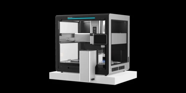
Traditionally bound by manual loading and processing, flow cytometry has recently experienced a jump in performance and throughput. The latest flow cytometers are designed to process 96- and 384-well microplates, detect dozens of colors and markers, and perform multi-parameter data acquisition. These state-of-the-art flow cytometers are suited to overcome key workflow bottlenecks within cell analysis and screening labs.
Cell analysis is challenging from a workflow and precision perspective. Streamlining the workflow using microplate handling and automated processing significantly lowers the burden. At the same time, increased throughput and multiplexing significantly improves precision, as more replicates and controls can be run simultaneously.
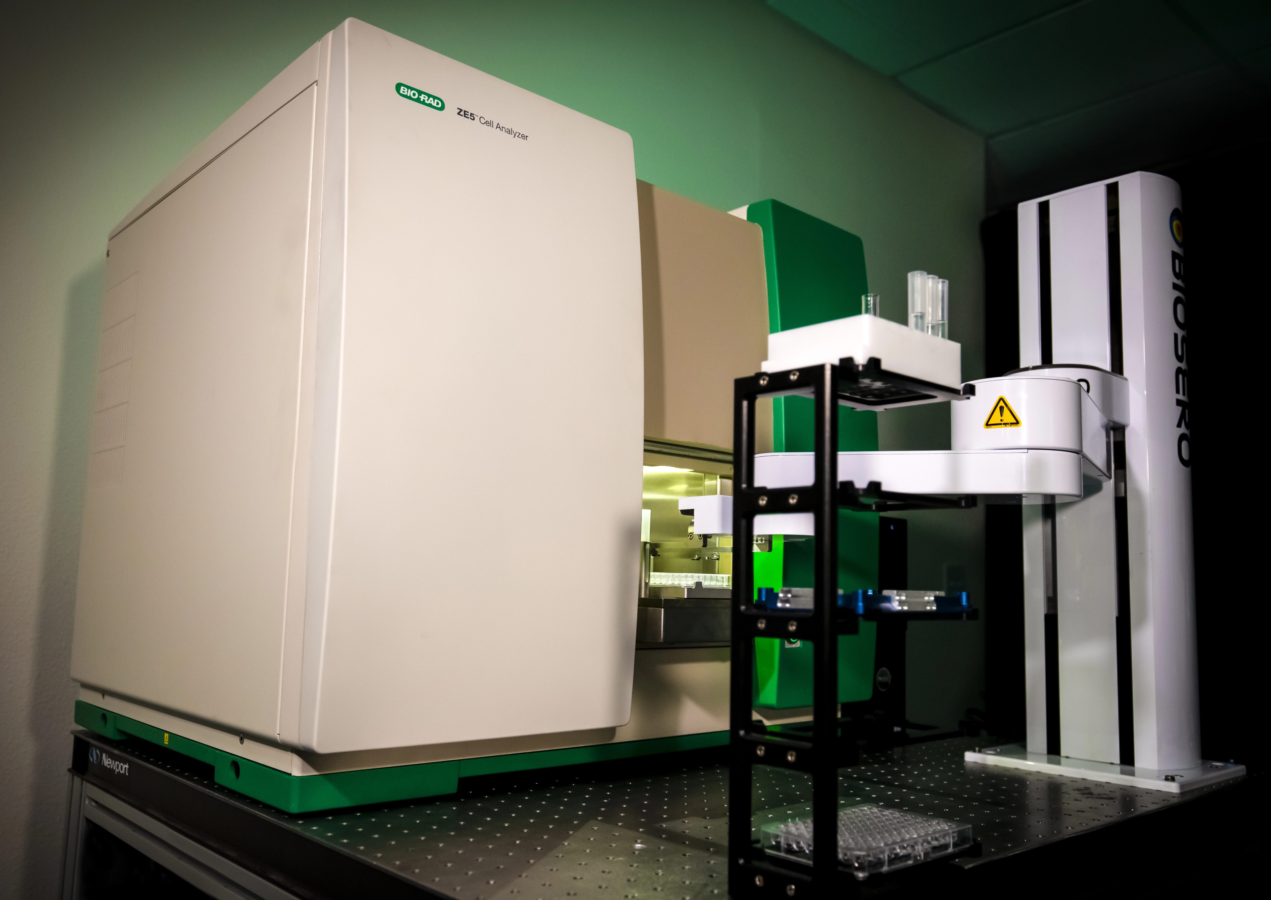 Bio-Rad now offers
a portfolio of automation-ready solutions to bring flow cytometry analysis up
to speed and in line with high-throughput screening (HTS) workflows. The Bio-Rad
ZE5 Cell Analyzer houses five lasers and 30 detectors for multi-parameter
detection. Fast sampling from 96- or 384-well plates, with speeds up to 100,000
events per second, equates to larger sample panels with faster
analysis times. Onboard diagnostics and high-capacity fluidics
tanks mean the system can be run 24/7 by a single operator.
The ZE5 can be easily integrated with lab robotics systems using a device-agnostic application programming interface (API). This makes it easy to program operations such as robotic microplate loading using scheduling software. Furthermore, the agnostic API allows communication between a wide range of automation providers and customized sample processing workflows.
Microplate-enabled automated 3D cell culture screening
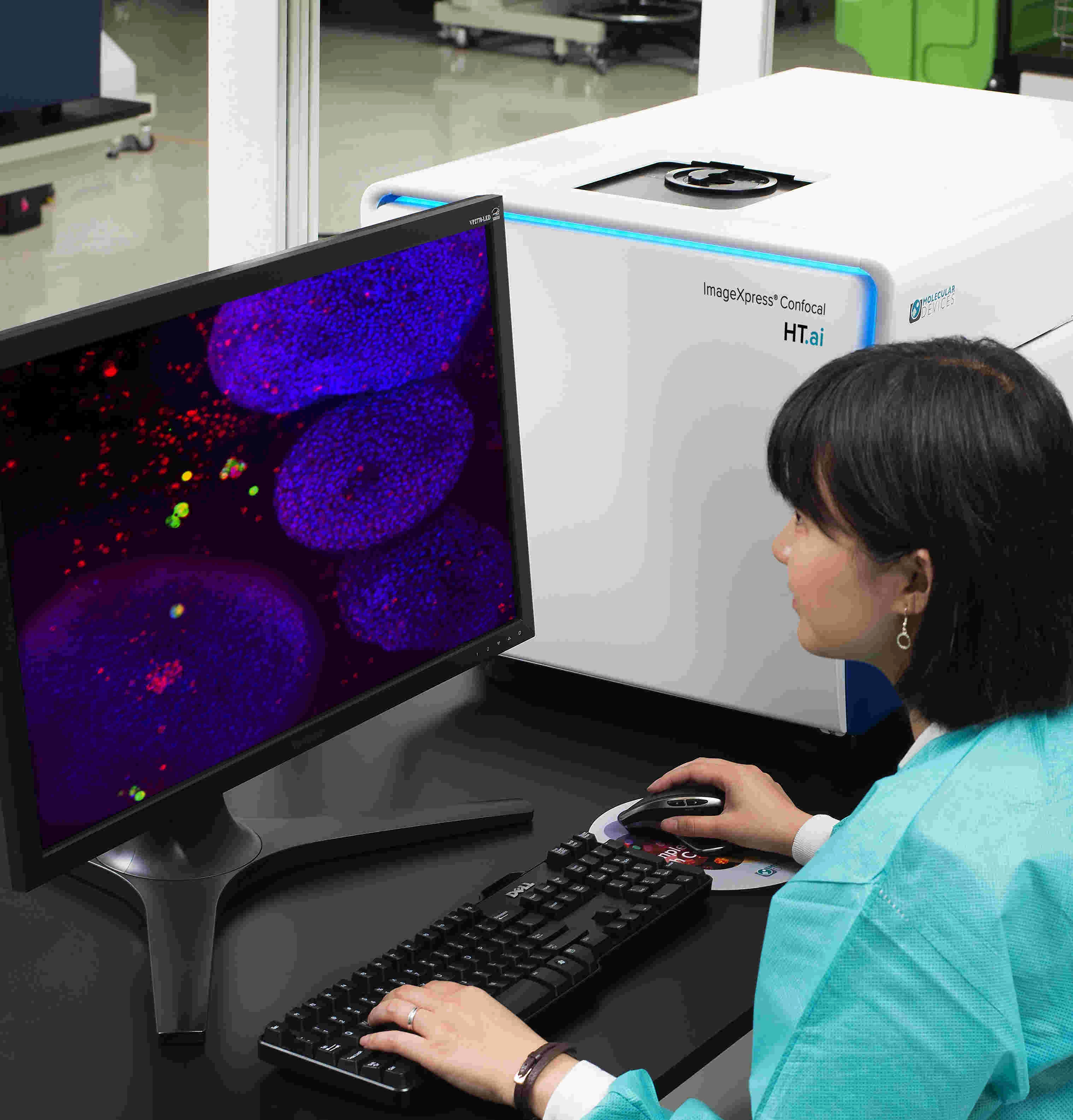 Microplate
technologies have proven essential in high-content imaging and high-throughput
screening applications. New technologies are now focusing on 3D cell culture
imaging coupled with HTS for drug discovery. As more complex biological 3D
models are created for drug research, more advanced imaging solutions must meet
the challenges.
Molecular Devices has created an Organoid Innovation Center to assist 3D cell culture and HTS-based drug discovery. This resource serves as an end-to-end solution for the entire organoid development process. Cell culture, treatment, incubation, imaging, analysis, and data processing are standardized to support unbiased biologically relevant results. The solutions also enable researchers to scaleup and automate HTS workflows.
The Organoid Innovation Center hosts a collaborative space, which allows researchers in the field to test automated workflows for culturing organoids and screening with guidance from in-house scientists. The overall goal of the center is to expand cell imaging by creating a fully-integrated solution that addresses all the challenges of complex biological screening – from sample preparation to data reporting.
Microplate-enabled live cell imaging
Traditional methods for imaging live cells involve removing cell cultures from the incubator and visualizing using a standard or fluorescence microscope. Sometimes samples are taken for cell staining and often cell counters are used to assess live or positive cell numbers.
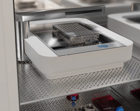 There are a
growing number of solutions to allow real-time imaging of cells without removal
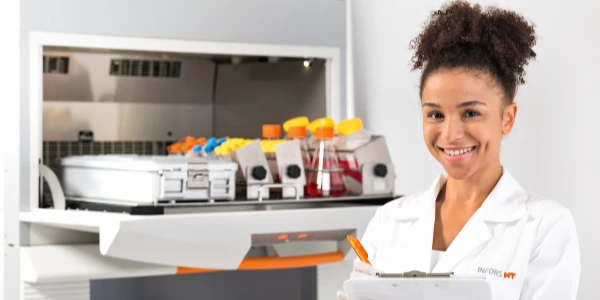
from the cell culture environment. A new product, the CytoSMART
Omni, is an automated live-cell analysis platform that allows continuous real-time
monitoring of multi-well cultures directly in the incubator.
The CytoSMART can scan all types of cell culture vessels including microplates, flasks, and custom microfluidic chips. Imaging functions include automated high-quality brightfield and/or fluorescence resolution of live cells. Data analysis software enables access to dynamic biological processes including cell growth, co-culture, transfection, cytotoxicity, and others. The imaging lens systematically scans the culture vessel, producing a complete survey of the landscape without disturbing the cells. Cells can be monitored, and data analysis can be performed, remotely providing a minimally invasive, automated solution for cell imaging analysis.
Summary
Microplate-enabled automated solutions are now supporting a range of challenging applications in the lab. Whether matching flow cytometry with HTS, assessing complex 3D cultures, or monitoring live cell events, emerging technologies aim to push the existing boundaries of cell analysis and imaging applications.
Visit the LabX Cell Culture Technology Application Page for further insight and product listings.
